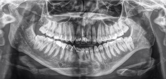
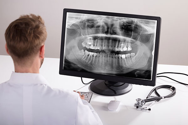
Dental x-rays are pictures that are taken of our teeth that show the underlying structures beneath the soft tissues, and in areas, we can’t visually see. X-rays are black and white images that show us the density of oral structures. On an x-ray, the lighter a structure is, the denser it is (called opaque), the darker a structure is, the less dense it is (called radiolucent.) Therefore enamel of teeth shows up very light, and gum tissue shows up very dark, almost not noticeable on an x-ray.
Why Are X-Rays Taken?
- Checking for cavities on biting surfaces, in-between the teeth and underneath the gums
- Checking for infections at the root (apex) of any of the teeth
- Monitoring the health and levels of the bone and periodontal ligament support
- Checking the angulation of tooth roots
- Checking for any abnormalities such as cysts
- Monitoring the health of past restorative work
- Checking wisdom teeth impaction
- Monitoring the health of the jaw joint
- Used for patients with dental braces (before and after)
- In kids, checking the eruption of baby teeth
- In kids, checking for extra or missing adult teeth under the gums

When Are X-rays Recommended?
X-rays are recommended based on an individual’s need. If there is an issue or concern, an x-ray will be needed to get a full picture of what is going on. Cavity check-up x-rays are recommended every 1-3 years based on cavity risk. Someone who has never had a cavity will be recommended less frequently, and someone who is cavity prone will be recommended more often.
Dental Considerations
X-rays are only taken when needed/ recommended. Most dental offices offer digital x-rays, which significantly reduce the radiation exposure. Lead aprons are used to cover the vital structures in the neck and torso. Regular check-up x-rays (a series of 4 x-rays) are around the same amount of radiation as the average background radiation in 1 day or the radiation from a 1-hour flight (very minimal!)

